Comparing spine curvatures
Keywords: spine, vertebral column, mouse, curvature, splines, statistical tests
Project partner: Dr. Alexis Ruiz Velez, Neuromuscular Research Laboratory at the Department of Biomedicine of the University of Basel
CeDA collaborator: Rodrigo C. G. Pena, Geoffrey Fucile
Repository: spine-curvatures
Context
Mutations in RYR1, the gene encoding the ryanodine receptor 1, the calcium channel of the sarcoplasmic reticulum, are the underlying cause of approximately 30% of all congenital myopathies. Patients usually present a range of phenotypes at a young age; these include developmental delays, muscle weakness, hypotonia and muscle stiffness that are primarily caused by defects in channel conductance or decreased amounts of ryanodine receptor protein, leading to less calcium being released from the sarcoplasmic reticulum. However, in addition to the skeletal muscle phenotype, many patients also present musculoskeletal defects including scoliosis, congenital hip dislocation, foot deformities, joint laxity etc. whose underlying causes have not been investigated.
Muscle spindles are an organ that is not involved in force generation, but act as mechanical sensors conveying information relating to muscle stretch and velocity of contraction to the central nervous system. This information is important not only for adjusting the tonic response of agonistic and antagonistic muscle groups, but also in modulating skeletal development. Indeed, patients with mutations in genes expressed in muscle spindles such as PIEZO2, exhibit musculoskeletal defects that are also present in patients harboring RYR1 mutations. We hypothesize that the skeletal deformities in patients harbouring RYR1 mutations are caused by malfunction of the intrafusal muscle fibres which make up the muscle spindles.
We hypothesize that defective muscle spindle function is an etiological factor of the musculoskeletal defects in patients with RYR1-related congenital myopathy. We will investigate this by studying the role of ryanodine receptor 1 in intrafusal muscle fibers in: (i) a mouse model we created knocked in for compound heterozygous RYR1 mutations and (ii) in a muscle-spindle specific RYR1 knock out mice.
Project objectives
The aim of this collaboration is to provide evidence for hypothesis above by comparing the spine curvatures in two groups of mice (control and transgenic).
Approaches
- Fit smooth curves (splines) to a point clouds sampled from a 3D scan of the mouse spines.
- Summarize the curvature within regions of interest in the vertebral column.
- Runs statistical tests to decide whether the curvature summaries come the same or different populations.
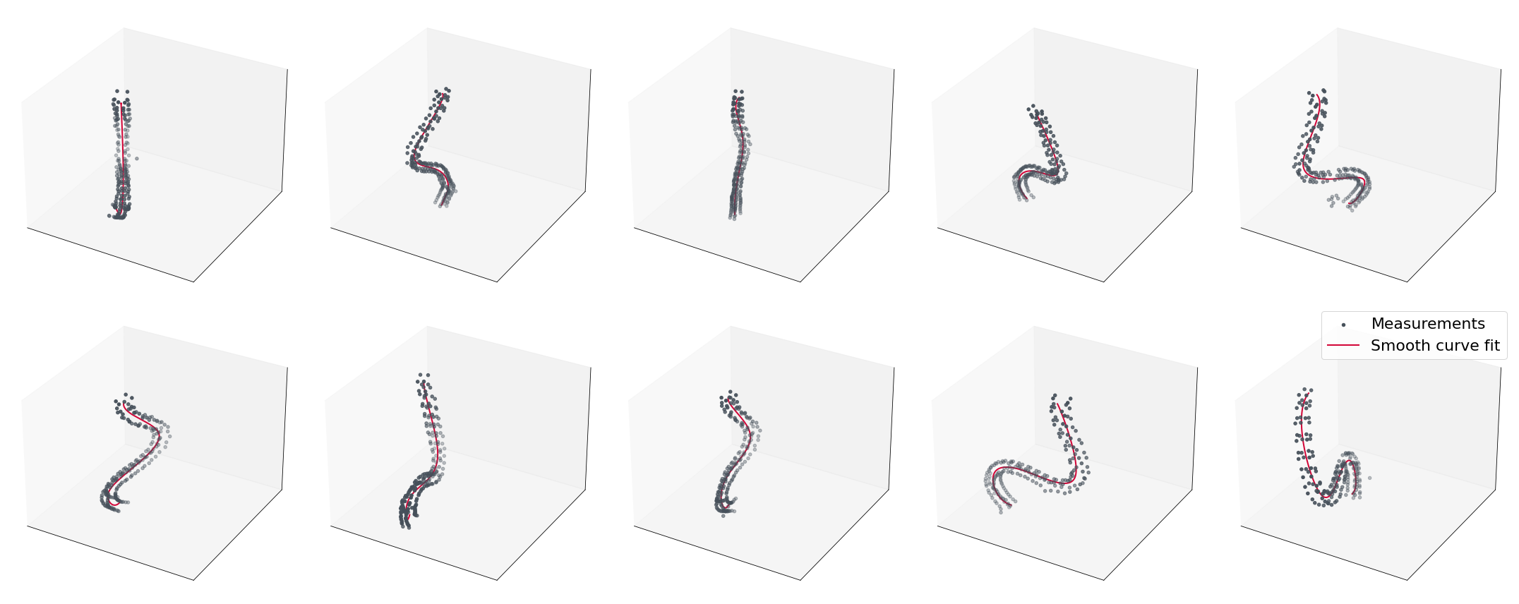
Figure 1. Spine point clouds (grey) and their corresponding fitted smooth curves (red). Each row represents a group of mice in the experiment.